
Photomicrography and Macrophotography Services
Science Photography & Education

Above Oxeye Daisy (Leucanthemum vulgare) at various magnifications including an aphid living inside the flower that was extracted and viewed by scanning electron microscopy showing the insect's compound eye and a single bacterium on the surface of the eye. These photos show the range of magnifications I can offer of various specimens.

Above is a wide variety of substances including wood, caffeine, bone, citric acid and starch grains viewed with a polarizing microscope.
If you or your business requires high magnification photographs for industry, medicine, forensic, education or personal use, including fiber optic, minerals, microchips, plankton, insects, Diatoms, Foraminifera, fossils, sand grains, coins, postage stamps, textiles, starch grains, botanical subjects, hair and other items contact me. I provide high quality images using specialized macrophotography and microscope equipment. By focus stacking multiple images I can increase the depth of field several fold and using stitching technology make images large enough for a billboard. I currently own nine different research quality microscopes providing a variety of specialized illumination techniques:
Phase contrast - for viewing living cells, aquatic organisms etc
Differential Interference Contrast - (DIC) viewing living cells and small organisms.
Polarization - for birefringent specimens, hair, wood, crystals, and minerals etc
Fluorescence - plants, microbes and other biological samples
Rheinberg lighting - a method to colourize variou sspecimens
Metallurgical microscopy - I can photograph opaque specimens and surfaces, metals etc
Darkfield illumination - aquatic organisms, bacteria, flagella etc
Brightfield illumination - stained slides and some aquatic organisms
Stereomicroscopy - offering magnificaitons from 1-200X
I can provide images with magnifications ranging from 0.5 X to 1500X using Light microscopy and even higher magnifications by renting time on a scanning electron microscope. I also provide HD movies and time lapse photography. Via an associate I can recommend someone that will cut stones and minerals 30 micron thick slides. I charge either by the image or hours of work at $60\hour. Educational classes cost the $60\hr and you can take images with you on a USB stick.
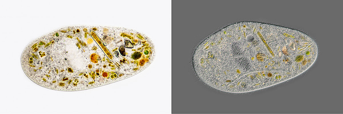
Comparison of a ciliate Frontia sp Left: bright field microscopy and Right: Differential interference microscopy 400X.
My light microscopes can resolve objects as small as 0.2 microns (0.0002 mm or 200 nm) and objects to about 20 nm with the scanning electron microscope. I provide image processing and optimization of the images. I can provide digital images on a CD\DVD, USB stick or portable hard drive. I have special tools for micromanipulation and can also prepare permanent microscope slides if required. I also provide video taken through the microscope and time lapse movies - see below. If you would like something photographed for artistic reasons, decorative purposes, post cards, posters or for educational purposes let me know.

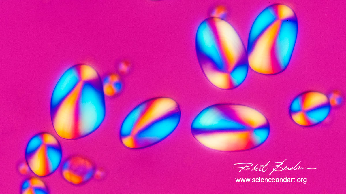
Citric acid crystals by Polarization Microscopy 100X
I am member of the University of Calgary Microscopy Group, an adjunct assistant professor in the Dept. of Cell Biology and I have access to Scanning electron microscopes. I am a trained scientist in the fields of Cell Biology, Neurobiology and Biochemistry with a passion for microscopy with over 50 years experience. I am also a professional Nature photographer and teacher and taught web design and photography at SAIT for over 20 years. My photographs have received international awards and have been published in magazines, science journals including numerous cover photos. I have also had images published by National Geographic and won awards from them. I have over 20 scientific publications in refereed sience journas, 5 book chapters, several books and hundreds of articles on microscopy - some for microscope companies like Motic. E.g. https://moticmicroscopes.com/blogs/articles.

Darkfield macrophotograph of a Trout Alevin 5X (Pulished in Scientific American).
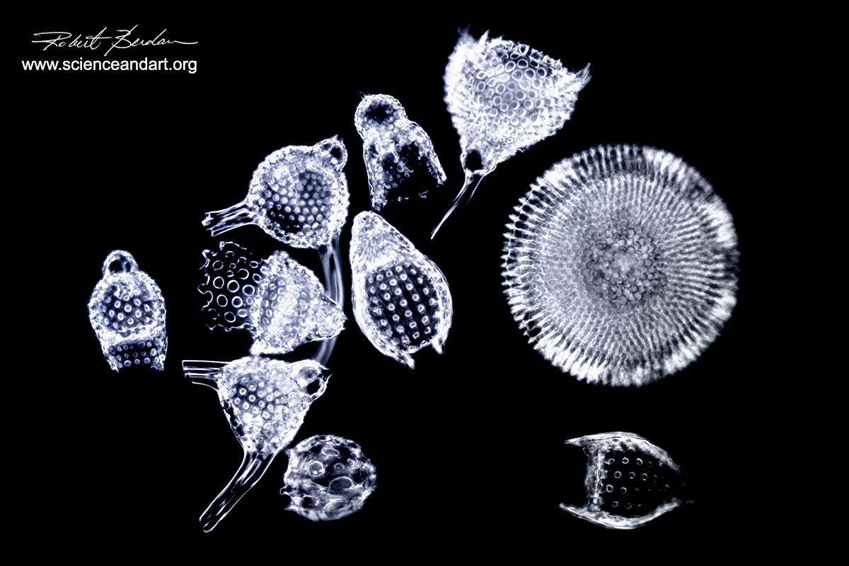
Radiolarians single cell plankton live in the ocean and form silica shells 400X Darkfield microscopy

Hexagonal symmetry of a snowflake 20X
Section through white Pine 400X Polarizing microscopy and full wave plate.

Leaf Section 100X Brightfield microscopy - panorama of images stitched together

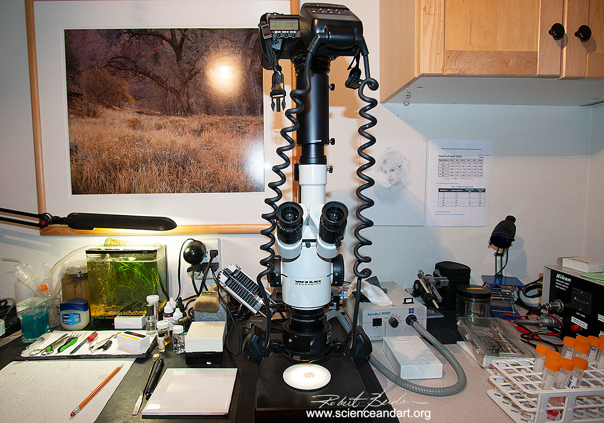
My private microscopy lab showing some of my microscopes.
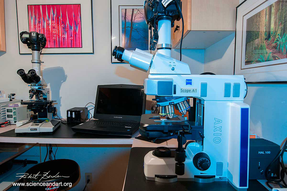
Left: Nikon Optiphot Polarizing\Fluorescence microscope Right: Zeiss Axioscope with DIC, Phase contrast, Polarizing and Darkfield illumination.
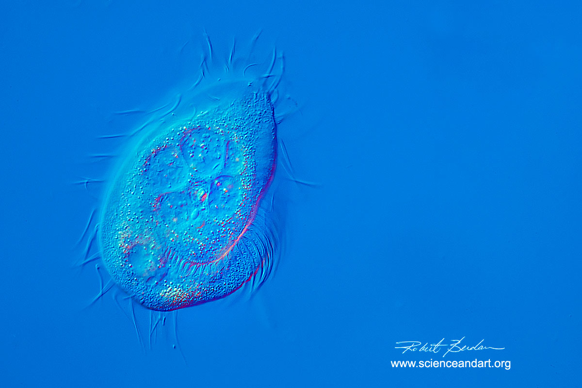
Phacodinium metchnikoffi a ciliate found in moss - Differential interference microscopy (DIC) 400X
Potato starch grains Polarized light with full wave plate 200X.

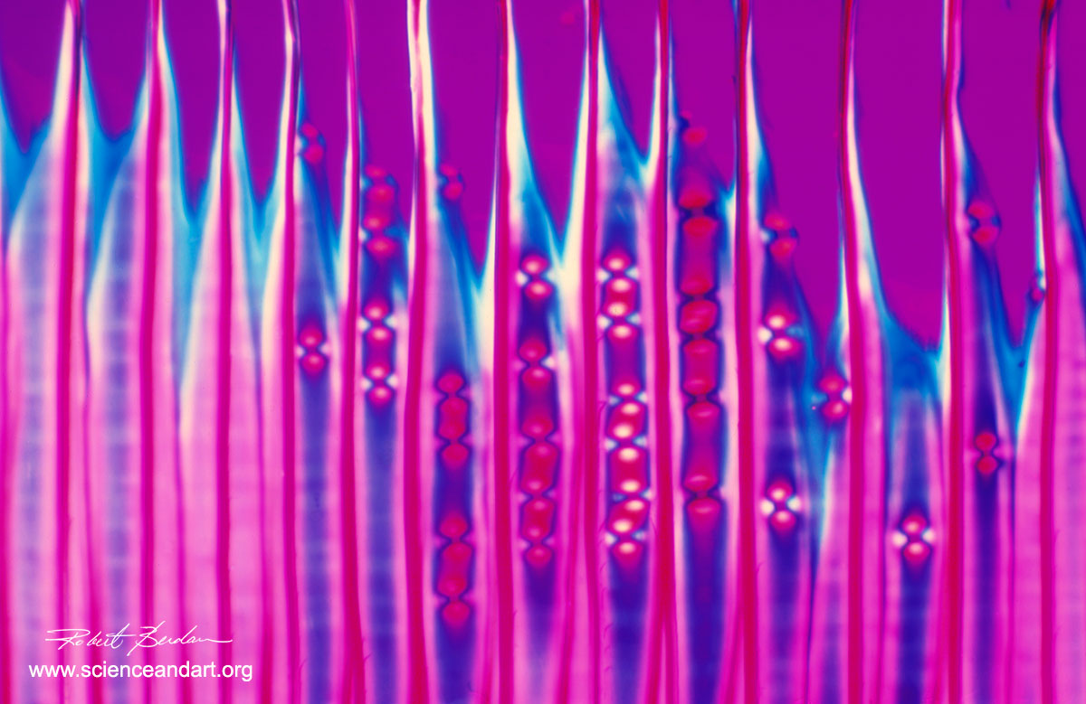
Above Citric Acid crystals in Polarized light - Abstract 100X
Wild Stereo microscope with a Canon 5D Mark II and electronic flash, ring-light and side lighting, capable of 1-50X magnification.
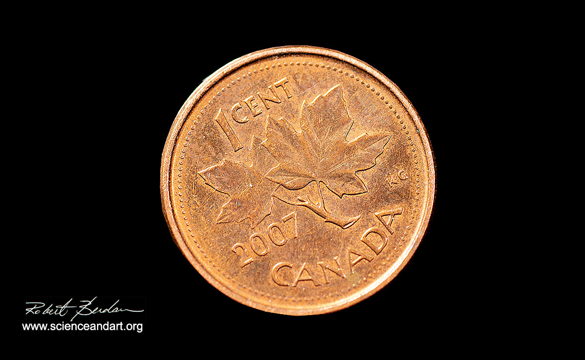
Canadian Penny photographed using the Wild Stereomicroscope.

Custom macro stand with Canon 80D and Canon MP 65-E lens with focus rail and high speed flash capable of 0.5 to 15X magnification.
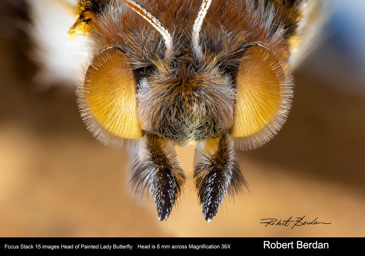
Painted Lady butterfly provided by an Alberta entomologist - 36X Focus stacked and photographed with the Canon camera and MP 65-E lens shown above.

Freshwater Diatoms photographed with the Olympus E microscope and Darkfield illumination. 400X
Scanning Electron Microscopy

Isolated neuron from the buccal ganglion of a Fresh water snail, Helisoma trivolvis, showing lipid vesicles below the plasma membrane by Scanning electron microscopy. False coloured 1000X

Scanning electron micrographs of single isolated neurons in culture for science publication.

Aphid low magnification using a Scanning electron microscope (SEM) 20X False coloured.
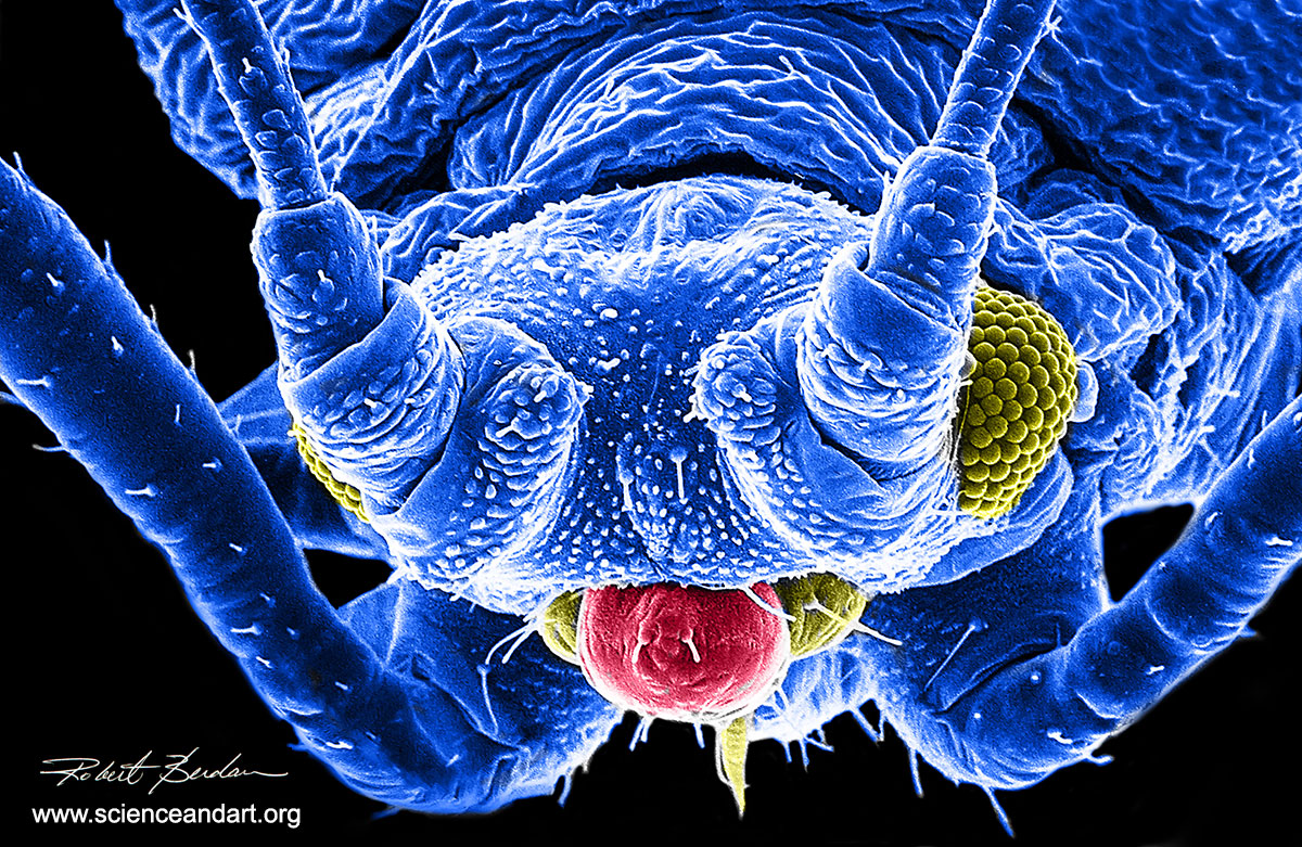
Head of an Aphid Scanning electron microscopy 200X - false coloured in Photoshop
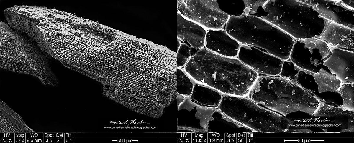
Bamboo charcoal showing individual cells by SEM
 Above is the tip of a mosquito proboscis by scanning electron microscopy.
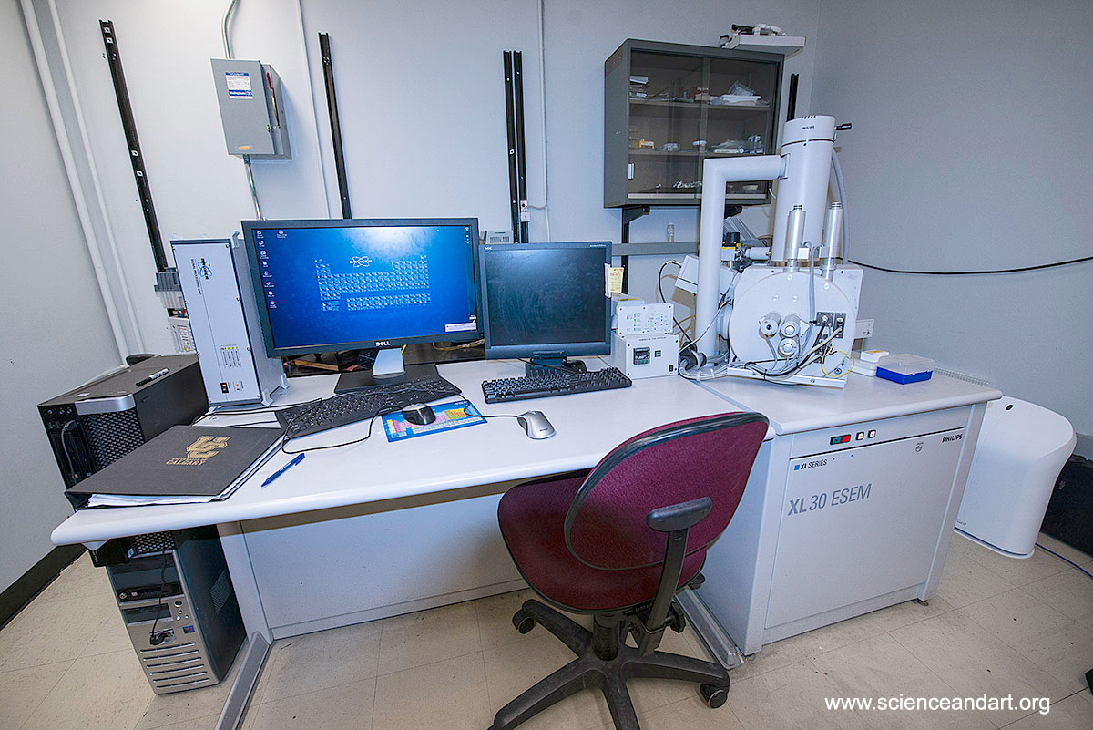
Scanning electron microscope at the University of Calgary - the microscope is available at an hourly rate.
Sections of Wood, Plants, Roofing Underlay, Vinyl Tiles and Polyester Filters Etc
I provide a variety of services which involve sectioning various materials and photographing them with my microscopes. I have sectioned roofing underlay, vinyl floor tiling, polyester filters used in the oil industry, plant and wood sections for the Calgary cience Center, fiber optic wires that were damaged. I own several microtomes, hand held and larger table top microtomes capable of sectioning a variety of materials. In addition I can also provide polarized light micrographs from various drinks like coffe, tea, sports drinks, pharmaceuticals, wine, beer and othe alcoholic drinks etc - see my article: Crystals Photographed with Polarization Microscopy: Water, Beer, Caffeine, Vitamins, Amino Acids and Human Tears.
 Sliding microtome for sectioning wood, vinyl, plants and other materials. I also uise a variety of smaller microtomes for cutting thin sections of filters,paper, plant tissues etc.
 Cross section of popular branch stained with safranin. Bright field microscopy 400X.

Crystals from a high energy drink viewed by Polarized light microscopy

Cross section of vinyl floor tile 15X

Vinyl floor tile cross section 25X

Viny floor tile cross section 50X


Below are a series of sections and Top views of Polyester filters

Above are cross sections and top views of various types of Polyester filters used in the oil industry.
Video of aquatic microorganisms created for Motic Microscopy - view above or on Youtube: https://youtu.be/Icvh83Mz7n4
Microscopy and Macrophotography Training and Equipment Rentals
I also offer training in all aspects of microscopy, macrophotography, and photomicrography for $60\hr or $299 per day (8 hours) in my studio. I offer 1-2 hours free training to students 12 years and older accompanied by an adult interested in learning about microscopy. Teachers or adults can receive microscopy traning for $60\hr and they may rent the use of some of my equipment in my studio afterwards for $40\hr to capture digital images using my cameras. I also provide private training to anyone interested in Photoshop and Focus stacking - contact me for more information or to book a training session. I am happy to recommend camera equipment, stereomicroscopes and light microscopes if you are considering purchasing one - just email me. For anyone visiting Calgary from afar there are a number of economical Air BNB locations nearby.
I sell some refurbished microscopes, I also clean, service and refurbish microscopes. For those looking for a new microscope I recommend Motic microscopes which offers professional equipment for about 1\4 the cost of the big 4 name brands (Zeiss, Leica, Nikon and Olympus) and offer high quality optics. Motic's head office for North America is in Vancouver. I reviewed and purchased their BA310 Polarizing microscope and looking to add phase contrast to this scope in the new year. In Calgary if you are looking for a stereomicroscope I recommend Doug Hayden owner of Quality microscopes. I own and use a Zeiss Stemi stereomicroscope from Doug. He sometimes has used equipment for sale as well.
To book my services or inquire about training please call 403 247-2457 or send an email to rberdan@scienceandart.org. I am available most weekdays 9 am to 9 pm, some evenings and weekends by apointment. If you are an advertising firm looking for unique images contact me for images, movies or specific services you might need for one of your clients.
To see more of my photomicrographs please visit my galleries and articles on my other web site www.canadiannaturephotographer.com. You can purchase my images for personal use as art, education or commercial use - see my prices.
Education
If you have a budding scientist in your family I would be happy to provide 1-2 hours free microscopy instruction in my studio for anyone between the ages of 12-17 under the supervision of a parent or guardian. I am happy to answer any questions you might have regarding microscopy and for an honorarium I would be willing to come to your school or organization in or around Calgary to provide a presentation on microscopy and photomicrography - The Microuniverse. Teachers or University students interested in learning more about microscopy are also welcome to visit me by appointment in my studio in Calgary, NW or take training.
My interest in microscopy began in Grade 6 and lead me into to a career in Cell biology and Neurobiology research and I would love to encourage others to take up microscopy as a hobby or a profession. The microworld is all around us waiting to be discovered - you can find interesting things in your backyard, nearby pond or in the soil. I have found interesting micro-organisms in rainwater from my evestrough and living in lichen and moss in my backyard.
See Article by Dr. Robert Berdan - The Micro-Universe - Microscopic Life
(403) 247-2457